ECHO

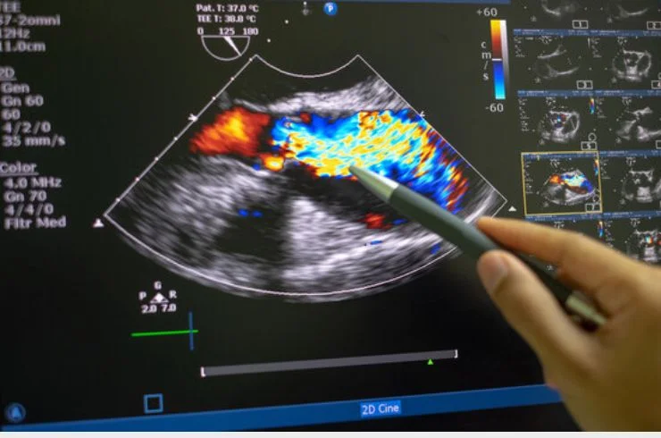
An Echocardiogram (ECHO) is a diagnostic test that uses sound waves (ultrasound) to create detailed images of the heart’s structure and function. It provides real-time, visual information about the heart’s chambers, valves, and blood flow, allowing doctors to assess the heart’s overall health and detect any abnormalities.
During the procedure, a gel is applied to the chest, and a small device called a transducer is moved over the skin. The transducer emits high-frequency sound waves that bounce off the heart and are then converted into images. These images can reveal the heart’s size, shape, and movement, as well as the condition of the heart valves and blood vessels.
Echocardiograms are commonly used to diagnose a wide range of heart conditions, including:
- Valvular diseases (e.g., valve stenosis or regurgitation)
- Heart failure and cardiomyopathy
- Congenital heart defects
- Pericardial effusion (fluid around the heart)
- Assessment of blood flow through the heart
This non-invasive procedure is critical for evaluating the heart’s pumping ability and guiding treatment for cardiovascular diseases. Different types of echocardiograms, such as stress ECHO or transesophageal ECHO, may be used for more detailed analysis in specific cases.

